
SegI restriction endonuclease from Sphingobium erigeronicola 46 recognizes the octanucleotide DNA sequence 5′-RTGC↓GCAY-3′ and is an isoschizomer of FspAI
D.A. Gonchar*, V.A. Chernukhin, M.A. Abdurashitov, V.S. Dedkov, N.A. Mikhnenkova, O.A. Belichenko, S.Kh. Degtyarev
SibEnzyme Ltd., Novosibirsk, Russia
* Corresponding author: D.A. Gonchar, SibEnzyme Ltd., 2/12 Timakova St., Novosibirsk, 630117, Russia; Tel.: +7 (383) 333-4991; Fax: +7 (383) 333-6853; E-mail: gonchar@sibenzyme.ru
ABSTRACT
We identified a bacterial strain, Sphingobium erigeronicola 46, that produces a novel rare-cutting restriction endonuclease, designated SegI. The enzyme recognizes the degenerate palindromic octanucleotide DNA sequence 5′-RTGC↓GCAY-3′ and cleaves both DNA strands in the center of the recognition site, as indicated by the arrow, generating blunt ends. SegI is therefore an isoschizomer of the previously described restriction endonuclease FspAI [1]. The recognition sequence was determined by analyzing the restriction patterns generated by SegI digestion of bacteriophage λ DNA (predigested with BssT1I), bacteriophage T7 DNA, and plasmid pBR322 DNA, followed by electrophoresis in a 0.8% agarose gel.
The producing strain was identified based on its morphological characteristics and sequence analysis of a 16S rRNA gene fragment. A SegI restriction endonuclease preparation with an activity of 5,000 U/mL was obtained through a three-step chromatographic purification procedure. The precise cleavage position within the recognition sequence was determined using a [³²P]-labeled oligonucleotide duplex. Optimal enzyme activity was observed at 37°C in SE buffer W (10 mM Tris-HCl, pH 8.5 at 25°C; 10 mM MgCl₂; 100 mM NaCl; 1 mM DTT).
Abbreviations: BSA, bovine serum albumin; DTT, dithiothreitol; T7 DNA, bacteriophage T7 DNA; λ DNA, bacteriophage lambda DNA; U, units of enzyme activity; Tris, tris(hydroxymethyl)aminomethane; PAGE, polyacrylamide gel electrophoresis; bp, base pairs; RE, restriction endonuclease; EDTA, ethylenediaminetetraacetic acid.
Keywords: enzyme-producing strain; enzyme purification; restriction endonuclease; isoschizomer.
DOI: 10.26213/3034-4301.2026.58.74.001
Citation:
Gonchar D.A., Chernukhin V.A., Abdurashitov M.A., Dedkov V.S., Mikhnenkova N.A., Belichenko O.A., Degtyarev S.Kh. (2026). SegI restriction endonuclease from Sphingobium erigeronicola 46 recognizes the octanucleotide DNA sequence 5′-RTGC↓GCAY-3′ and is an isoschizomer of FspAI. DNA-Recognizing Enzymes, 2026(5). https://doi.org/10.26213/3034-4301.2026.58.74.001
This article is licensed under a Creative Commons Attribution-NonCommercial-NoDerivatives 4.0 International License.
INTRODUCTION
Type II restriction endonucleases cleave double-stranded DNA within or near their recognition sites, generating either sticky or blunt ends [2]. These properties have made them indispensable tools in molecular biology and genetic engineering. In this study, we describe a novel rare-cutting restriction endonuclease, SegI, which recognizes the degenerate palindromic octanucleotide DNA sequence 5′-RTGCGCAY-3′, where R represents a purine and Y a pyrimidine, and cleaves both DNA strands at the center of the recognition site, generating blunt ends.
MATERIALS AND METHODS
Reagents from Sigma-Aldrich (USA), Fisher (Germany), Panreac (Spain), Dia-M, and Helicon (Russia) were used throughout this study. The following chromatography media were employed for enzyme purification: phosphocellulose P-11 and heparin-Sepharose (Sigma-Aldrich, USA), and hydroxyapatite (Bio-Rad, USA). Components of bacterial growth media were obtained from Organotechnie (France).
Restriction endonucleases, T4 polynucleotide kinase, bacteriophage λ and T7 DNA preparations, plasmid pBR322, DNA molecular weight markers, SE enzyme dilution buffer B100, and SE reaction buffers B, G, O, W, Y, and ROSE were obtained from SibEnzyme Ltd. (Russia).
The morphological, physiological, and biochemical characteristics of the bacterial strain were examined using standard methods [3]. The genus of the microorganism was identified based on Bergey’s Manual [4], in which this genus was not yet represented, and sequence analysis of a PCR-amplified fragment of the 16S rRNA gene.
To determine the position of phosphodiester bond cleavage, a [³²P]-labeled DNA duplex containing the recognition site of the target enzyme was used. The oligonucleotides were synthesized by BIOSSET Ltd. (Russia) and radiolabeled using T4 polynucleotide kinase. The enzymatic cleavage products of the labeled oligonucleotides were separated by electrophoresis in a 20% denaturing polyacrylamide gel containing 8 M urea.
Cultivation of the Enzyme-Producing Strain
The enzyme-producing strain was grown in 500-mL Erlenmeyer flasks containing 300 mL of LM broth (1% tryptone, 0.5% yeast extract, 0.5% NaCl, 0.05% MgCl₂, and 0.001% thiamine) at 28°C with shaking for 48 h until the cultures reached saturation. Cells were harvested by centrifugation in a Beckman J2-MI centrifuge (USA) using a JA-10 rotor at 8,000 rpm for 20 min and stored at −20°C.
Purification of SegI Restriction Endonuclease
Purification conditions and buffers. All purification steps were performed at 4°C using solutions based on the following buffers:
- Buffer A: 10 mM Tris-HCl, pH 7.6; 0.1 mM EDTA; 7 mM β-mercaptoethanol.
- Buffer B: 10 mM potassium phosphate, pH 7.5; 0.1 mM EDTA; 7 mM β-mercaptoethanol.
Cell extraction. A total of 27 g of cell biomass was suspended in 125 mL of buffer A supplemented with 0.2 M NaCl, 1 mg/mL lysozyme, and 0.1 mM PMSF (phenylmethylsulfonyl fluoride, a protease inhibitor). After stirring for 1 h on a magnetic stirrer, the suspension in a 150-mL glass beaker was cooled in an ice bath. Cells were then disrupted by sonication at maximum power (22–24 μm amplitude) using a Soniprep 150 sonicator (MSE, UK) equipped with a 2-cm-diameter probe. Six 1-min pulses were applied at 1-min intervals. The resulting cell lysate was clarified by centrifugation in a Beckman J2-MI centrifuge (USA) using a JA-20 rotor at 15,000 rpm for 30 min.
Phosphocellulose P-11 chromatography. A 30-mL phosphocellulose P-11 column was washed with 60 mL of buffer A containing 0.2 M NaCl. Bound proteins were eluted with a 500-mL linear gradient of 0.3–0.7 M NaCl in buffer A. Fifty 10-mL fractions were collected. Fractions exhibiting the highest SegI activity were pooled and loaded onto a 3-mL hydroxyapatite column pre-equilibrated with buffer B.
Hydroxyapatite chromatography. The column was washed with 10 mL of buffer B. The enzyme was eluted with a 100-mL linear gradient of 10–150 mM potassium phosphate buffer (pH 7.5) containing 0.05 M NaCl. Forty 2.5-mL fractions were collected. Fractions exhibiting the highest enzyme activity were pooled and applied to a 5-mL heparin-Sepharose column pre-equilibrated with buffer A containing 0.2 M NaCl.
Heparin-Sepharose chromatography. The column was washed with 10 mL of buffer A containing 0.2 M NaCl. Bound proteins were eluted with a 120-mL linear gradient of 0.2–0.6 M NaCl in buffer A. Fifty 2.5-mL fractions were collected.
Concentration and Storage of the Enzyme Preparation
Fractions exhibiting the highest SegI activity were pooled, and BSA was added to a final concentration of 0.2 mg/mL. The pooled fractions were dialyzed against 200 mL of concentration buffer (10 mM Tris-HCl, pH 7.6; 0.1 mM EDTA; 7 mM β-mercaptoethanol; 0.2 M NaCl; 50% glycerol) with continuous stirring for 16 h. The resulting SegI preparation was stored at −20°C.
Assay of SegI Restriction Endonuclease Activity
Bacteriophage T7 DNA, which contains two SegI recognition sites, was used as the substrate for enzyme activity assays. To monitor restriction endonuclease activity during chromatographic purification, 1-μL aliquots of individual fractions were added to 20-μL reaction mixtures containing 1 μg of T7 DNA in SE buffer W (10 mM Tris-HCl, pH 8.5 at 25°C; 10 mM MgCl₂; 100 mM NaCl; 1 mM DTT). Reaction mixtures were incubated at 37°C for 15 min.
The digestion products were separated by electrophoresis in a 0.8% agarose gel using Tris-acetate buffer (50 mM Tris-acetate, pH 8.0; 20 mM sodium acetate; 2 mM EDTA) at 150 V. Following ethidium bromide staining, the gel was photographed under UV illumination.
One unit of SegI activity was defined as the minimum amount of enzyme required to completely digest 1 μg of bacteriophage T7 DNA in SE buffer W at 37°C within 1 h in a total reaction volume of 50 μL.
To determine the DNA cleavage position of SegI, a 22-bp oligonucleotide duplex, Seg1/Seg2, was used. The duplex was 5′-end labeled with [³²P] and contained the SegI recognition site as well as recognition sites for control restriction endonucleases. It was formed by annealing two synthetic oligodeoxyribonucleotides with the following sequences:
Seg1: 5′-GGTTTTCATGCGCATGTTTTGG-3′
Seg2: 5′-CCAAAACATGCGCATGAAAACC-3′
RESULTS AND DISCUSSION
Characterization and Taxonomic Identification of the Enzyme-Producing Strain
The enzyme-producing strain was isolated from a freshwater sample. When grown on LM agar at 24°C for 3 days, the strain formed circular, convex, shiny, translucent yellow colonies up to 2 mm in diameter. The cells were rod-shaped (0.6 × 2–3 μm), motile, Gram-negative, catalase-positive, and oxidase-negative. The strain grew under aerobic conditions at temperatures ranging from 8 to 30°C but showed no growth at 37°C.
The G+C content of the genomic DNA was determined to be 65 ± 5% using the method described in [5]. For taxonomic identification, the 16S rRNA gene was amplified by PCR using the primers 5′-AGAGTTTGATCMTGGCTCA-3′ and 5′-TACGGYTACCTTGTTACGACT-3′, which are modified versions of the fD1 and rP1 primers [6]. Two fragments of the 16S rRNA gene, 511 and 626 bp in length, were sequenced (Fig. 1).
Figure 1. Nucleotide sequences of two fragments of the 16S rRNA gene from the enzyme-producing strain.
   1 TTAGATCCTG GCTCAGTCGT CTTCGGTGAG CCGGACAGTT GGATTGCTGG CTCGTGGGAA
  61 TATGGCCTTG GGTTCGGAAT AACTTCGGGA AACTGAAGCT AATACCGGAT GATGACGAAA
 121 GTCCAAAGAT TTATCGCCCA AGGATGAGCC CGCGTAGGAT TAGCTAGTTG GTGGGGTAAA
 181 GGCCTACCAA GGCGACGATC CTTAGCTGGT CTGAGAGGAT GATCAGCCAC ACTGGGACTG
 241 AGACACGGCC CAGACTCCTA CGGGAGGCAG CAGTAGGGAA TATTGGACAA TGGGGGCAAC
 301 CCTGATCCAG CAATGCCGCG TGAGTGATGA AGGCCTTAGG GTTGTAAAGC TCTTTTACCC
 361 GAGATGATAA TGACAGTATC GGGAGAATAA GCCCCGGCTA ACTCCGTGCC AGCAGCCGCG
 421 GTAATACGGA GGGGCTAGCG TTGTCGGAAT TACTGGGCTA AAGCGCACGT AGGCGGCGAT
 481 TAAGTCAGAG TGAAGCCGGG CTCACCCCGG A


   1 AACGATGATA CTAGCTGCCG GGCACATGGT GTTTCGGTGG CGCAGCTAAC GCATTAAGTT
  61 ATCCGCCTGG GGAGTACGGT CGCAAGATTA AAACTCAAAG GAATTGACGG GGGCCTGCAC
 121 AAGCGGTGGA GCATGTGGTT TAATTCGAAG CAACGCGCAG AACCTTACCA ACGTTTGACA
 181 TCCCTATCGC GGTTACCAGA GATGGTTTCC TTCAGTTCGG CTGGATAGGT GACAGGTGCT
 241 GCATGGCTGT CGTCAGCTCG TGTCGTGAGA TGTTGGGTTA AGTCCCGCAA CGAGCGCAAC
 301 CCTCGCCTTT AGTTGCCAGC ATTAAGTTGG GTACTCTAAA GGAACCGCCG GTGATAAGCC
 361 GGAGGAAGGT GGGGATGACG TCAAGTCCTC ATGGCCCTTA CGCGTTGGGC TACACACGTG
 421 CTACAATGGC GACTACAGTG GGCAGCCACT CCGCGAGGAG GAGCTAATCT CCAAAAGTCG
 481 TCTCAGTTCG GATCGTTCTC TGCAACTCGA GAGCGTGAAG GCGGAATCGC TAGTAATCGC
 541 GGATCAGCAT GCCGCGGTGA ATACGTTCCC AGGCCTTGTA CACACCGCCC GTCACACCAT
 601 GGGAACAAAT CACCCGAAGG SGTTGA
Based on sequence analysis of the 16S rRNA gene fragments, the enzyme-producing strain was identified as Sphingobium erigeronicola, a member of the family Sphingobiaceae. Taxonomic identification was performed using Nucleotide BLAST [7] and the NCBI Taxonomy Browser [8]. The microbiological characteristics of the strain were consistent with those reported for this species [9]. The strain was designated Sphingobium erigeronicola 46, and the restriction endonuclease isolated from it was named SegI in accordance with the established nomenclature [10].
Biomass Production and Enzyme Isolation
Cultivation of Sphingobium erigeronicola 46 yielded 7.5 ± 0.5 g of cell biomass per liter of culture. A total of 27 g of biomass, exhibiting a SegI activity of 10,000 U/g, was obtained from 3.6 L of culture and stored at −20°C. Following the three-step chromatographic purification procedure described above, 4 mL of purified SegI preparation was obtained and stored at −20°C.
To determine the substrate specificity of the new restriction endonuclease, we examined its ability to cleave three different DNA substrates. Figure 2 shows the restriction patterns generated by SegI digestion of bacteriophage λ DNA predigested with BssT1I (λ/BssT1I), bacteriophage T7 DNA, and plasmid pBR322 DNA. Digestion reactions were performed in 50-μL mixtures containing 1 μg of substrate DNA and 1 μL of SegI preparation in SE buffer W. The reaction mixtures were incubated at 37°C for 1 h.
Figure 2. Electrophoretic analysis of various DNA substrates digested with the restriction endonuclease SegI.
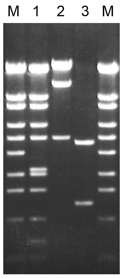
Lanes:
M, λ/BssT1I DNA molecular weight marker (SibEnzyme);
1, λ/BssT1I DNA + 1 μL SegI preparation;
2, bacteriophage T7 DNA + 1 μL SegI preparation;
3, plasmid pBR322 DNA + 1 μL SegI preparation.
Cleavage products were separated by electrophoresis in a 0.8% agarose gel.
Comparison of the cleavage patterns generated by the new enzyme on the DNA substrates described above (Fig. 2) with theoretically predicted restriction patterns for previously characterized restriction endonucleases indicated that SegI is an isoschizomer of the previously described enzyme FspAI [1]. This enzyme recognizes the degenerate palindromic octanucleotide DNA sequence 5′-RTGCGCAY-3′, with two recognition sites present in each of the three DNA substrates: bacteriophage λ DNA, bacteriophage T7 DNA, and plasmid pBR322 DNA.
Determination of the Optimal Reaction Buffer and Temperature for SegI
To determine the optimal reaction buffer for SegI, digestion reactions were performed in 50-μL mixtures containing one of six standard SE restriction endonuclease buffers (B, G, O, W, Y, or ROSE), 1 μg of bacteriophage T7 DNA, and either 0.5 or 1 μL of the SegI preparation diluted 5-fold in SE buffer B100. Reaction mixtures were incubated at 37°C for 1 h. The results are shown in Fig. 3.
Figure 3. SegI activity on bacteriophage T7 DNA at 37°C in six SE reaction buffers (B, G, O, W, Y, and ROSE). For each buffer, either 0.5 or 1 μL of the SegI preparation diluted 5-fold was added to the reaction mixture. Digestion products were analyzed by electrophoresis in a 1% agarose gel.
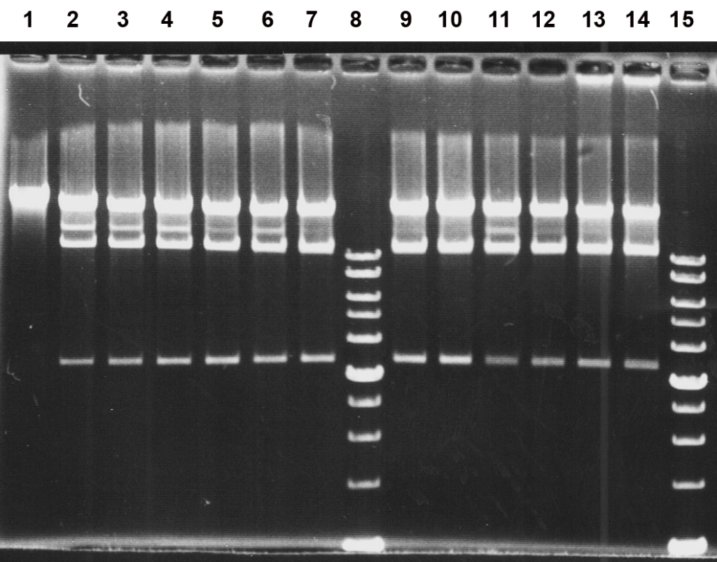
Lanes:
1, undigested bacteriophage T7 DNA;
2–3, SE buffer B;
4–5, SE buffer G;
6–7, SE buffer O;
9–10, SE buffer W;
11–12, SE buffer Y;
13–14, SE buffer ROSE;
8 and 15, SibEnzyme 1 kb DNA ladder.
As shown in Fig. 3, SE buffer W provides optimal conditions for SegI activity. Complete digestion of the substrate DNA was achieved with 1 μL of the enzyme preparation diluted 5-fold (lane 10). Under these conditions, T7 DNA (39,935 bp) was cleaved into three fragments of 25,453, 11,071, and 3,412 bp. Partial digestion resulted in additional fragments, including a 14,483-bp fragment (11,071 + 3,412 bp) migrating between the 25,453- and 11,071-bp fragments, and a 28,865-bp fragment (25,453 + 3,412 bp) that was not resolved from the 25,453-bp fragment.
Complete digestion of T7 DNA was also observed in SE buffer ROSE (lane 14). The relative activities of SegI in the six standard SE reaction buffers at 37°C are summarized in Table 1.
Table 1. Activity of SegI restriction endonuclease in standard SE reaction buffers at 37°C.
| SE Buffer | B | G | O | W | Y | ROSE |
|---|---|---|---|---|---|---|
| SegI Activity, % | 10–25 | 25–50 | 50–75 | 100 | 50–75 | 100 |
To determine the optimal reaction temperature for SegI, digestion reactions were performed in 50-μL mixtures containing SE buffer W, 1 μg of bacteriophage T7 DNA, and 0.5, 1, or 2 μL of the SegI preparation diluted 5-fold in SE buffer B100. Reaction mixtures were incubated for 1 h at 25, 30, 37, or 55°C. As a control at each temperature, T7 DNA was digested in SE buffer W using 1 μL of undiluted SegI preparation. The results are shown in Fig. 4.
Figure 4. Activity of SegI restriction endonuclease on bacteriophage T7 DNA in SE buffer W at different temperatures. Digestion products were analyzed by electrophoresis in a 1% agarose gel.
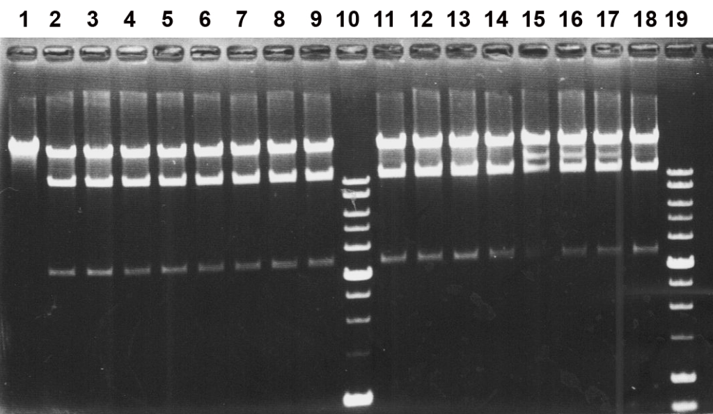
Lanes:
1, undigested bacteriophage T7 DNA;
2, 6, 11, 15, T7 DNA + 0.5 μL of SegI preparation diluted 5-fold;
3, 7, 12, 16, T7 DNA + 1 μL of SegI preparation diluted 5-fold;
4, 8, 13, 17, T7 DNA + 2 μL of SegI preparation diluted 5-fold;
5, 9, 14, 18, T7 DNA + 1 μL of undiluted SegI preparation;
2–5, incubation at 25°C;
6–9, incubation at 30°C;
11–14, incubation at 37°C;
15–18, incubation at 55°C;
10 and 19, SibEnzyme 1 kb DNA ladder.
As shown in Fig. 4, complete digestion of the substrate DNA at all enzyme dilutions was observed at 37°C (lanes 11–14), indicating that this temperature is optimal for SegI activity. The enzyme also exhibited high activity at 25 and 30°C (lanes 2–5 and 6–9, respectively). In contrast, at 55°C, complete digestion was no longer achieved with the diluted enzyme preparation (lanes 15–17), although SegI retained at least 50% of its activity. Complete inactivation of SegI in the reaction mixture was achieved by heating at 65°C for 20 min (data not shown).
Based on the results presented in Figs. 3 and 4, one unit of SegI activity was defined as the minimum amount of enzyme required to completely digest 1 μg of bacteriophage T7 DNA in a 50-μL reaction mixture containing SE buffer W at 37°C within 1 h.
Figure 5 shows the electrophoretic analysis of SegI activity under optimal reaction conditions (37°C in SE buffer W).
Figure 5. Determination of SegI activity under optimal reaction conditions. Reaction mixtures (50 μL) contained 1 μg of bacteriophage T7 DNA in SE buffer W and 0.5, 1, or 2 μL of the SegI preparation diluted 5-fold in SE buffer B100. The mixtures were incubated at 37°C for 1 h, and digestion products were analyzed by electrophoresis in a 1% agarose gel.
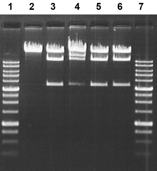
Lanes:
2, undigested bacteriophage T7 DNA;
3, bacteriophage T7 DNA + 1 μL of undiluted SegI preparation (control);
4, bacteriophage T7 DNA + 0.5 μL of SegI preparation diluted 5-fold;
5, bacteriophage T7 DNA + 1 μL of SegI preparation diluted 5-fold;
6, bacteriophage T7 DNA + 2 μL of SegI preparation diluted 5-fold;
1 and 7, SibEnzyme 1 kb DNA ladder.
As shown in Fig. 5, complete digestion of the substrate DNA was achieved when either 1 or 2 μL of the 5-fold diluted SegI preparation was added to the reaction mixture (lanes 5 and 6, respectively). Thus, the activity of the purified enzyme preparation was determined to be at least 5 U/μL, corresponding to 5,000 U/mL.
Determination of the DNA Cleavage Position of SegI
To determine the precise DNA cleavage position of the new enzyme, we used a 22-bp oligonucleotide duplex, Seg1*/Seg2, radiolabeled at the 5′ end (the labeled strand is indicated by an asterisk), containing the SegI recognition site (underlined):
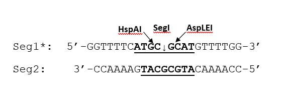
The duplex was digested separately with SegI and three control restriction endonucleases: SmiMI (5′-CAYNN↓NNRTG-3′), HspAI (5′-G↓CGC-3′), and AspLEI (5′-GCG↓C-3′). The cleavage positions of these enzymes within the duplex are indicated by arrows in the sequence diagram above.
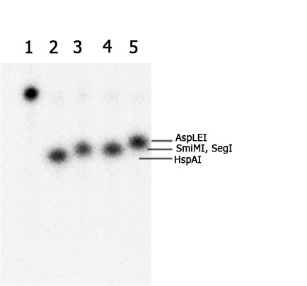
Cleavage reactions were performed in 10-μL mixtures containing 1 pmol of the Seg1*/Seg2 duplex and 1 μL of the respective enzyme in SE buffer W. Reaction mixtures were incubated at 37°C for 1 h. The cleavage products were separated by electrophoresis in a 20% denaturing polyacrylamide gel containing 8 M urea, using Tris–borate buffer. Figure 6 shows the resulting gel autoradiogram.
Figure 6. Determination of the SegI cleavage position. Cleavage products were separated by electrophoresis in a 20% denaturing polyacrylamide gel containing 8 M urea.
Lanes:
1, undigested Seg1*/Seg2 duplex;
2, Seg1*/Seg2 duplex + 1 μL HspAI;
3, Seg1*/Seg2 duplex + 1 μL SmiMI;
4, Seg1*/Seg2 duplex + 1 μL SegI;
5, Seg1*/Seg2 duplex + 1 μL AspLEI.
The results presented in Fig. 6 demonstrate that the cleavage position of SegI coincides with that of SmiMI (5′-CATGC↓GCATG-3′) (lanes 3 and 4). The SegI cleavage position differs by one nucleotide from those of both AspLEI, which generates a slightly longer cleavage product (lane 5), and HspAI, which produces a shorter fragment (lane 2). Thus, SegI cleaves both DNA strands symmetrically at the center of its recognition sequence, 5′-CATGC↓GCATG-3′/3′-GTACG↓CGTAC-5′, generating blunt ends.
Most restriction endonuclease prototypes identified to date recognize DNA sequences of 4–6 bp [2]. Only approximately 15 prototypes with 8-bp recognition sites have been described. These include enzymes recognizing perfect palindromes, interrupted palindromes, and palindromic sequences containing degenerate positions.
The enzyme described in this study recognizes a degenerate palindrome. Restriction endonucleases with this type of recognition sequence are relatively rare, with only a few prototypes reported, including FspAI, SgrAI, and PspXI. It is possible that restriction endonucleases recognizing such sequences evolved from enzymes with palindromic hexanucleotide recognition sites.
This hypothesis is supported by the observation that the hexanucleotide sequence 5′-TGCGCA-3′, recognized by MstI, Acc16I, and FspI, forms part of the FspAI and SegI recognition sites. MstI and its isoschizomers cleave this sequence at the same position relative to the central hexanucleotide, generating blunt ends identical to those produced by FspAI and SegI.
The novel restriction endonuclease SegI described in this study may prove useful for generating large DNA fragments in genetic engineering, molecular biology, biotechnology, and DNA diagnostics.
REFERENCES
- Kesminiene A, Maneliene Z, Vitkute J, Petrusyte M, Janulaitis A. (2001). FspAI, a unique type II restriction endonuclease that recognizes the octanucleotide sequence 5′-RTGC^GCAY-3′. Nucleic Acids Research. 29:e120.
- Pingoud A, Fuxreiter M, Pingoud V, Wende W. (2005). Type II restriction endonucleases: structure and mechanism. Cellular and Molecular Life Sciences. 62:685–707. https://doi.org/10.1007/s00018-004-4513-1
- Egorov NS, ed. (1995). Manual for Practical Classes in Microbiology. Moscow, Russia. (In Russian).
- Holt JG, et al., eds. (1997). Bergey’s Manual of Determinative Bacteriology. 9th ed. Russian translation in 2 volumes, edited by G.A. Zavarzin. Moscow, Russia. (In Russian).
- Dedkov VS. (2004). Determination of G+C content in bacterial DNA using restriction endonucleases. Biotekhnologiya (Biotechnology). 4:77–82. (In Russian).
- Weisburg WG, Barns SM, Pelletier DA, Lane DJ. (1991). 16S ribosomal DNA amplification for phylogenetic study. Journal of Bacteriology. 173:697–703. https://doi.org/10.1128/jb.173.2.697-703.1991
- National Center for Biotechnology Information (NCBI). Nucleotide BLAST. https://blast.ncbi.nlm.nih.gov
- National Center for Biotechnology Information (NCBI). NCBI Taxonomy Browser. https://www.ncbi.nlm.nih.gov/Taxonomy/
- Jeong CR, Kim SB. (2026). Description of Sphingobium wonjuense sp. nov. isolated from mountain soil and Sphingobium erigeronicola sp. nov. isolated from plant root. International Journal of Systematic and Evolutionary Microbiology. 76(3).
- Smith HO, Nathans D. (1973). A suggested nomenclature for bacterial host modification and restriction systems and their enzymes. Journal of Molecular Biology. 81:419–423.